Medical Imaging · Digital Pathology
Pathology artifact segmentation
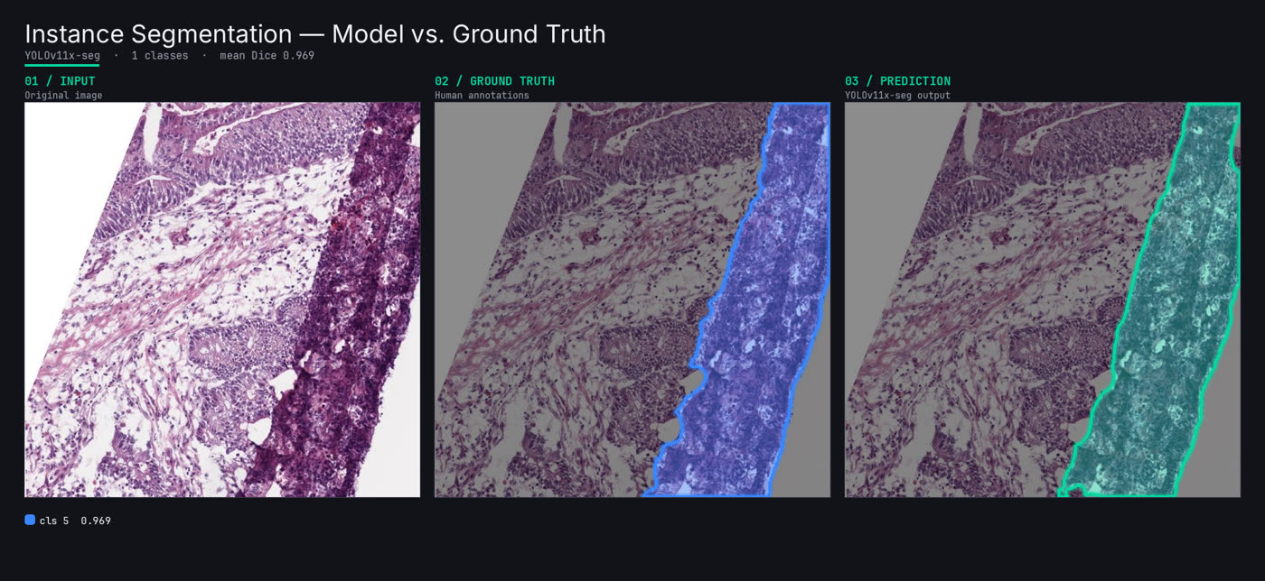
An instance-segmentation model that flags 11 artifact types in whole-slide pathology images — dust, pen, folds, air bubbles, tissue scratches, focus blur — rebuilt to beat the client's existing baseline.
The challenge
An underperforming baseline
The client already had an artifact model — its accuracy simply wasn't good enough to rely on.
11 subtle artifact types
Dust, pen, folds, air bubbles, and scratches look alike and are easily confused with tissue.
Severe class imbalance
Some artifacts appear in hundreds of instances, others in only one or two.
The approach
Dataset audit & cleanup
Consolidated 356 images and 423 instances across the 11 artifact classes, fixing label noise.
YOLO11x-seg model
Replaced the baseline with a 62M-parameter YOLO11x instance-segmentation backbone.
Imbalance-aware training
Tuned training to lift long-tail classes without sacrificing the dominant ones.
Per-class error analysis
Per-class Dice, COCO mask mAP, and a confusion matrix to expose where classes bled together.
Outcomes
Mask mAP@50
YOLO11x-seg across all 11 classes (mask mAP@50–95 0.48).
Best-class Dice
Clean artifacts like debris segmented near-perfectly.
Full artifact taxonomy
Every slide-artifact type covered by one model.